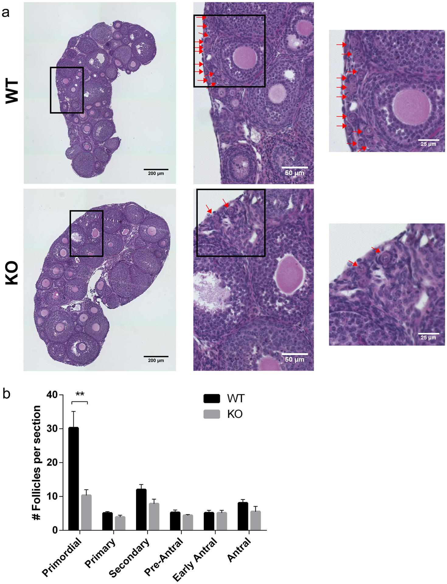
Fig. 3. Reduced number of primordial follicles in the absence of SIRT7.

(a) Representative images of hematoxylin/eosin-stained ovarian sections from 3-week-old wild-type (WT) and Sirt7−/− (KO) females. Red arrows denote primordial follicles in all panels. The boxes indicate the regions of zoom. (b) Quantification of follicle types from the ovaries represented in (a). Follicle numbers were quantified for each ovary and reported as the average number of each type of follicles per section. Graph represents the mean ±SEM (n=4 animals/genotype). ** P < 0.01
