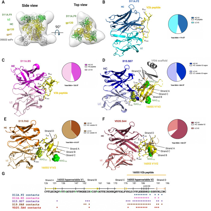
Fig 2. Vaccine-elicited antibodies recognize the V2 region of 16055 strain.
(A) nsEM 3D reconstruction with low resolution crystal structure of D11A.F9 Fab (Heavy chain, dark green; Light chain, light green) and 35022 scFv (gray) in complex with 16055 NFL (gp120, yellow; gp41, light brown) shown in two different views. (B, C) Structures of D11A.F2 Fab (Heavy chain, sky blue; Light chain, cyan) and D11A.B5 Fab (Heavy chain, magenta; Light chain, light pink) bound to the V2b peptide (yellow). (D) Structures of D15.SD7 (Heavy chain, blue: Light chain, light blue), (E) D19.PA8 (Heavy chain, orange; Light chain, light orange) and (F) VD20.5A4 (Heavy chain, raspberry; Light chain, light raspberry) Fabs in complex with the 16055 V1V2-1FD6 scaffold. (B, C, D, E, F) Interacting residues are shown in sticks and glycans in green. Pie charts summarize the buried surface area (BSA) of the V2b and V1V2-1FD6. (G) Sequence of 16055 V1V2 highlighting the V2b peptide used for crystallization, the location of the V1, V2 and strands. Residues that contact the Mabs (within 5Å) are shown with asterisks underneath the sequence. N-linked glycosylation sites are shown in green.

