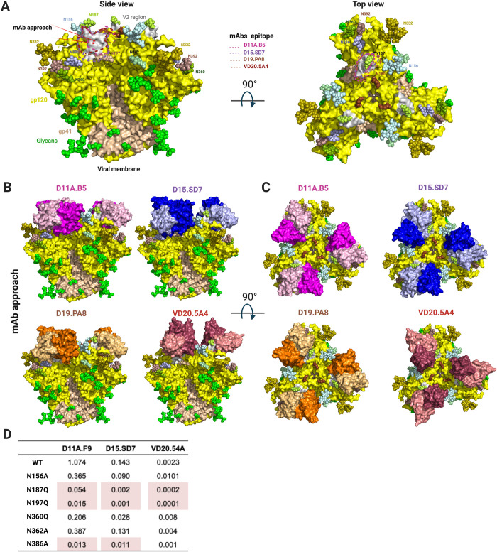
Fig 7. NHP Autologous tier 2 neutralizing antibodies target a hole in the HIV-1 glycan shield.
(A) Side and top view surface representation of 16055 NFL (PDB:5UM8) with gp120 shown in yellow, V2 region in grey, gp41 in wheat and glycans shown in green spheres or color-coded and labeled. Arrows indicate mAbs’ angle of approach. Epitopes targeted by the NHP mAbs are highlighted. (B) Side view and (C) Top view superpositions of the structures of D11A.B5, D15.SD7, D19.PA8 and VD20.5A4 onto the 16055 NFL trimer, showing how they access the glycan hole. Trimer and mAbs are shown in surface representation. Trimer is color coded as in (A) and mAbs as in Fig 3. (D) Effect of glycan removal surrounding the epitope on neutralization potency. Neutralization IC50 values (μg/ml) shown with >10-fold differences highlighted in red.

