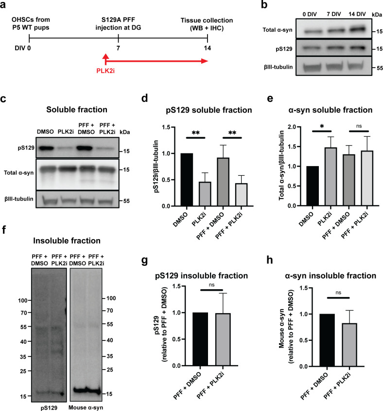
Fig 1. PLK2i treatment reduces S129-phosphorylated α-syn in the soluble fraction but not the insoluble fraction of organotypic slices.
a) Workflow of the experiment. PLK2 inhibition is started 24 hours before S129A PFF injection and continued until tissue collection at 7 dpi. The timeline is depicted as days in vitro (DIV). b) Immunoblotting of OHSCs from wild type (C57Bl6) pups, showing expression of endogenous α-syn and pS129 α-syn at 0, 7 and 14 days in culture, demonstrating the presence of a basal level of physiological RIPA-soluble pS129 α-syn in the slices. c) Immunoblotting of the RIPA-soluble fraction of PLK2i- or DMSO-treated slices ± PFFs for pS129 and total α-syn, demonstrating reduction of pS129 in PLK2i-treated slices in a PFF-independent manner. d & e) Quantification of immunoblots for pS129 (d) and total α-syn (e) in the RIPA-soluble fraction. Band values relative to βIII-tubulin were normalized to DMSO/non-injected group and represent mean ± SD. n = 3 independent experiments of 8 to 10 slices per group in each experiment. Statistical comparisons were performed using one-way ANOVA with Fisher’s LSD as post hoc test, * p < 0.05, ** p < 0.01. P-values for pS129 (d) were 0.004 (DMSO vs. PLK2i) and 0.007 (PFF+DMSO vs. PFF+PLK2i). P-values for total α-syn (e) were 0.0498 (DMSO vs. PLK2i) and 0.6607 (PFF+DMSO vs. PFF+PLK2i). f) Immunoblotting of the RIPA-insoluble fraction of PFF-injected slices ± PLK2i shows that PLK2 inhibition does not affect generation of aggregates or S129-phosphorylation of α-syn in the insoluble fraction. Representative blot from 3 independent experiments. Molecular size markers in kDa are indicated. g & h) Quantification of immunoblots for pS129, p-value = 0.9604 (g), and mouse α-syn, p-value = 0.3421 (h), in the RIPA-insoluble fraction. n = 3 independent experiments of 8 to 10 slices per group in each experiment. Statistical comparisons were performed using Welch’s test.

