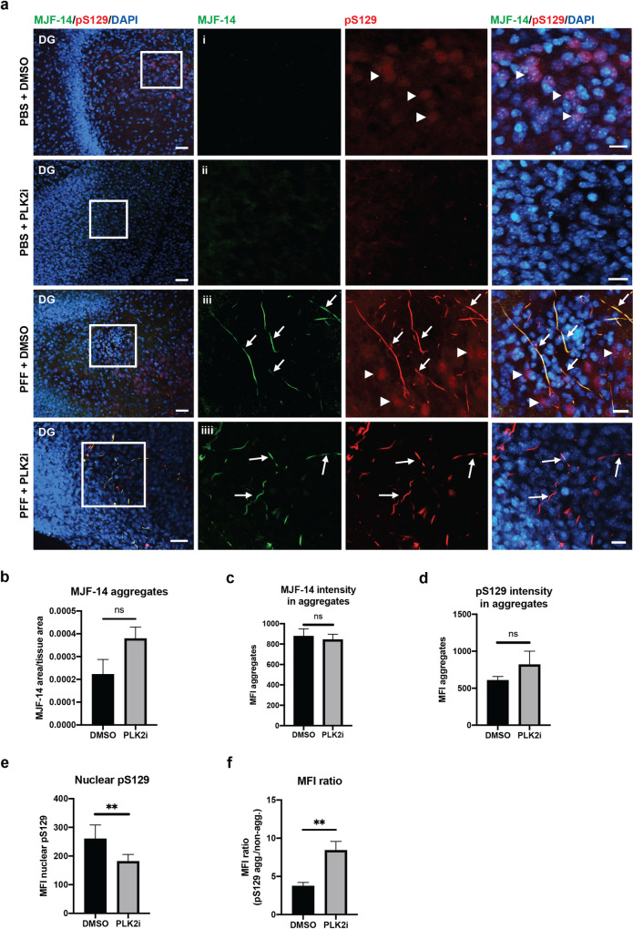
Fig 2. PLK2i treatment of OHSCs does not affect generation of pS129 positive aggregates, but reduces the nuclear pS129 intensity.
a) Representative images from the dentate gyrus region (DG) of organotypic slices injected with PBS or S129A PFFs and treated with DMSO or PLK2i. Slices were stained with MJF-14, pS129 (11A5), and DAPI, scale bar = 50 μm. i. The magnified region (white boxed area) shows nuclear pS129-signal that co-localizes with DAPI (arrowheads). ii. The nuclear pS129-staining was removed following PLK2i treatment. iii. S129A PFF-induced α-syn aggregates, phosphorylated at S129, are detected with MJF-14 and pS129 antibodies (arrows). pS129-staining of non-aggregated α-syn is also seen, predominantly located in the nuclei (arrowheads). iiii. PLK2i treatment shows no effect on the S129-phosphorylation of aggregated α-syn (arrows) but effectively reduces the nuclear pS129-staining. Scale bars = 20 μm. b) Quantification of the amount of aggregation (aggregate area normalized to tissue area), defined by MJF-14 staining (p-value = 0.09). c-d) Quantification of the mean fluorescence intensity of α-syn aggregates detected with MJF-14 (c, p-value = 0.712) and aggregate-specific pS129 that overlaps with MJF-14 (d, p-value = 0.214). e) Quantification of nuclear (non-aggregate related) pS129, illustrating a significant decrease in mean fluorescence intensity following PLK2i-treatment (p-value = 0.0087). f) Relative ratio of aggregate-specific pS129/nuclear pS129 in PFF-injected slices shows an increase of ratio of the signals of aggregates/background following PLK2i treatment, due to reduction of the non-aggregate-related nuclear pS129-signal (p-value = 0.009). Bars represent mean ± SD of 3 independent experiments with 8 to 10 slices/experiment. Treatments were compared using an unpaired Student’s T-test.

