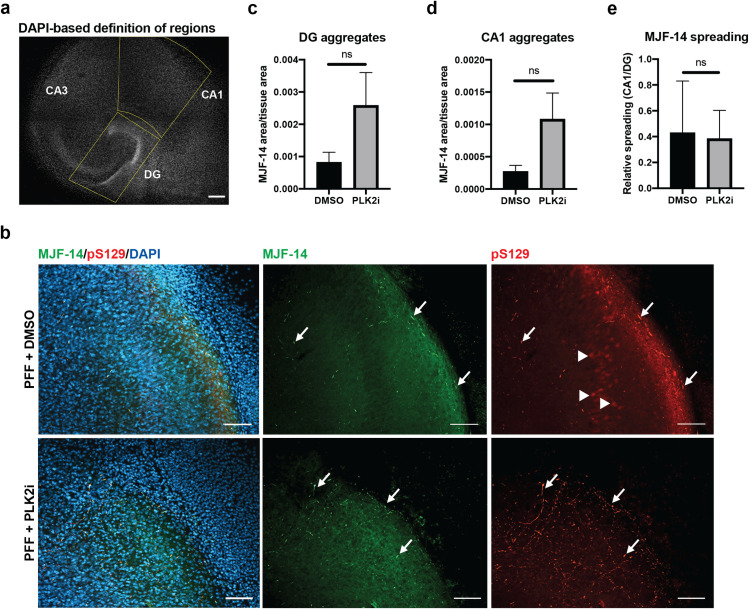
Fig 3. Inter-neuronal spreading of a-syn aggregate pathology from the DG to the CA1 region occurs independently of PLK2 inhibition.
a) Segmentation of DG and CA1 regions in organotypic hippocampal slices labelled with DAPI, shown in gray scale. The DG is bounded manually by a box, and the CA1 regions is afterwards defined by extrapolation of one side of the DG box and a diagonal line through the box. Scale bar = 200 μm. b) Immunostaining of S129A PFF-injected OHSCs showing aggregated α-syn at the CA1 region 7 dpi detected by both MJF-14 and pS129. PLK2i-treatment reduces the nuclear pS129-signal at CA1 that overlaps with DAPI (arrowheads) but has no influence on the PFF-induced aggregate-specific pS129-signal that overlaps with MJF-14-signal (arrows). Scale bars = 50 μm. c-d) Quantification of α-syn aggregate area (MJF-14 area normalized to tissue area) at the DG (c) and CA1 region (d) shows no significant effects on aggregation at either region following PLK2i treatment. P-values 0.1502 (c) and 0.1048 (d) using an unpaired Welch’s T test. e) Aggregate levels at the CA1 region were normalized to aggregate levels in DG of the same slice to address the relative spreading of S129A PFF-induced α-syn aggregates, based on MJF-14 staining. PLK2i treatment did not affect relative spreading of MJF14-positive pathology (p-value = 0.829 using an unpaired Welch’s T test). Bars represent mean ± SD of 5–6 slices per group. Images are representative of three independent experiments.

