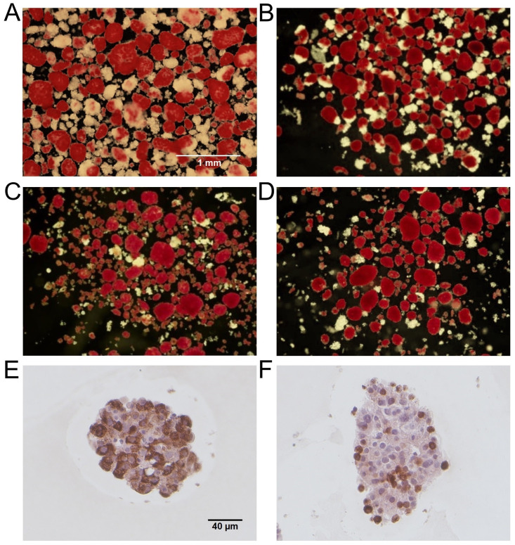
Fig 1. Representative human islet preparations.
Representative dithizone-stained images of human islets used in the study with purity of 75% (A, n = 2), 80% (B, n = 3), 90% (C, n = 5), and 95% (D, n = 4). Dithizone-stained endocrine cells (red) and non-dithizone-stained exocrine cells (white) are shown. Scale bar represents 1mm. Human islets stained for insulin (E) and glucagon (F), counterstained with Harris’ hematoxylin and eosin. Scale bar represents 40µm.

