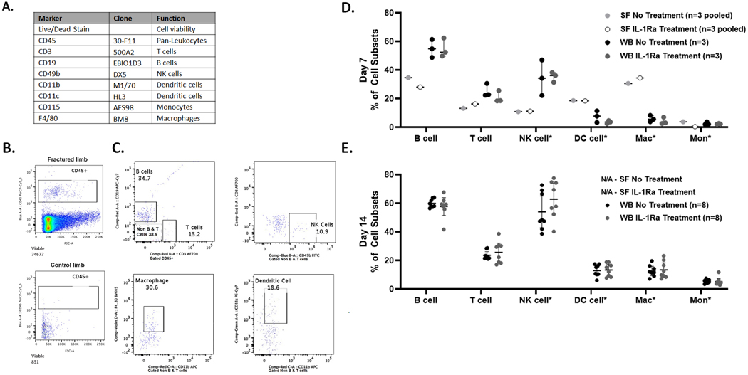
Figure 3.
Immune cell phenotyping in synovial fluid and whole blood from mouse model of closed articular fracture via polychromatic flow cytometry A) Panel of antibodies for immune cell phenotyping. B) Comparison of CD45+ leukocytes in pooled synovial fluid from 3 joints at 7 days post-fracture of fractured (74,677 events) and control (851 events) limbs with no treatment. C) Representative example of detection of B cells, T cells, NK cells, macrophages and dendritic cells (DC) in the synovial fluid from untreated fractured limbs, gated on CD45+ for T cells and B cells and *Non-B and T cells for remaining cell subsets. D) Immune cell subsets at Day 7 post-fracture in synovial fluid (SF) and whole blood (WB) for both no treatment and IL-1Ra treatment (mean ± 95% CI). E) Immune cell subsets at Day 14 post-fracture in whole blood (mean ± 95% CI). Synovial fluid data Not Available (N/A) from pooled synovial fluid from 4 joints (n=2 per group) as cell count, determined by detectable events, was too low for subset analysis.

