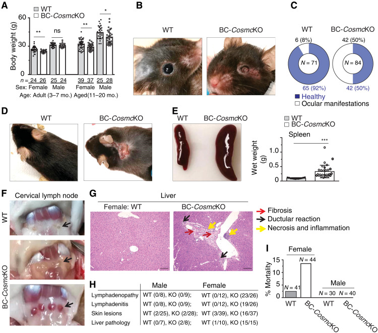
Fig. 1. AID-like pathological features in BC-CosmcKO mice.
Obvious phenotypic abnormalities of aged [11 to 20 months (mo.)] female and male mice from both BC-CosmcKO and WT littermate controls were documented (A to F). Body weight of both adult (3 to 7 months) and aged mice from both genders is shown (A): adult female: WT 26.75 ± 3.37, BC-CosmcKO 24.58 ± 1.26; adult male: WT 31.25 ± 2.15, BC-CosmcKO 30.57 ± 2.09; aged female: WT 32.26 ± 5.06, BC-CosmcKO 29.14 ± 3.27; aged male: WT 44.48 ± 6.92, BC-CosmcKO 39.61 ± 7.68. Each symbol (black square, WT; open circle, BC-CosmcKO) represents an individual mouse body weight, graphed as means ± 1 SD. Ocular manifestations (B and C) and dermatitis (D). Spleens (E) and cervical lymph nodes (F) of aged female mice. Representative images from both WT and BC-CosmcKO mice are shown in (B) to (F); n = 12 for WT and n = 26 for BC-CosmcKO (E). (G) Liver damage in aged female Cosmc mutant mice. Liver sections were stained with hematoxylin and eosin (H&E). Yellow arrows indicate necrosis and inflammation. Black arrows indicate ductular reaction. Red arrows indicate fibrosis. (H) Summary of examined animals in (D), (F), and (G). Liver H&E images were acquired at ×20 magnification. Scale bars, 100 μm. (I) Bar graphs represent mortality summary of both BC-CosmcKO and WT littermate controls. Each symbol (black square, WT; open circle, BC-CosmcKO) represents an individual mouse, graphed as means ± 1 SEM. ns, not significant. Unpaired two-tailed Student’s t tests were performed to determine statistical significance, *P < 0.05, **P < 0.01, and ***P < 0.001. Photo credit: Junwei Zeng, Beth Israel Deaconess Medical Center.

