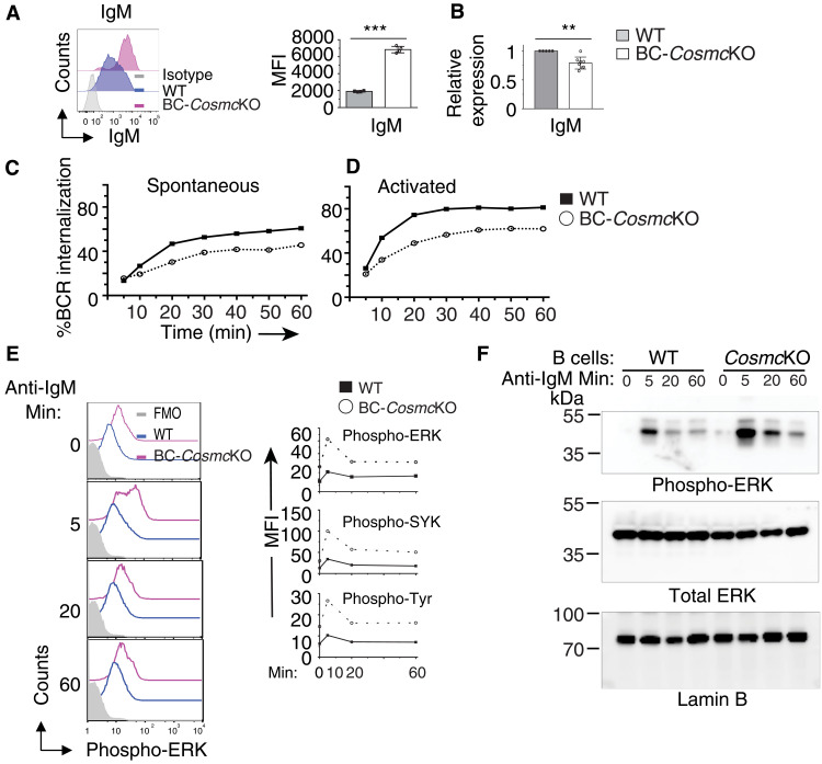
Fig. 5. Cosmc deficiency leads to impaired BCR internalization and enhanced BCR signaling.
(A) Representative histogram plot of IgM surface expression on B220+ B cells of both WT and BC-CosmcKO mice. Right: MFI of IgM on B cells. Results are representative of three independent experiments with at least three animals in each group. (B) Transcript level of IgM in purified B cells of both WT and BC-CosmcKO mice. n = 5 for WT and n = 8 for BC-CosmcKO. (C) Spontaneous IgM internalization by WT and BC-CosmcKO B cells incubated for indicated periods of time as measured by biotinylated Fab′ anti-IgM antibody. (D) Ligand-activated IgM internalization by WT and BC-CosmcKO B cells incubated for indicated periods of time as measured by biotinylated F(Ab′)2 anti-IgM antibody. Experiments in (C) and (D) were repeated four times with similar results. Each symbol (black square, WT; open circle, BC-CosmcKO) represents an individual mouse. Graphed as means ± 1 SEM. Young (2 to 4 months old) male mice were used in (A) to (F). Unpaired two-tailed Student’s t tests were performed to determine statistical significance, **P < 0.01 and ***P < 0.001. (E) Phospho-flow cytometry analysis of WT and BC-CosmcKO B cells stimulated with anti-IgM antibody for indicated periods of time, assessing the intensity of phosphorylated SYK, ERK1/2, and total cellular proteins (pan-tyrosine). Right: MFI of indicated proteins. Experiments were repeated four times with similar results. (F) Phosphorylated ERK1/2 Western immunoblotting of WT and BC-CosmcKO B cells stimulated with anti-IgM antibody (10 μg/ml) for indicated periods of time. Experiments were repeated two times with similar results.

