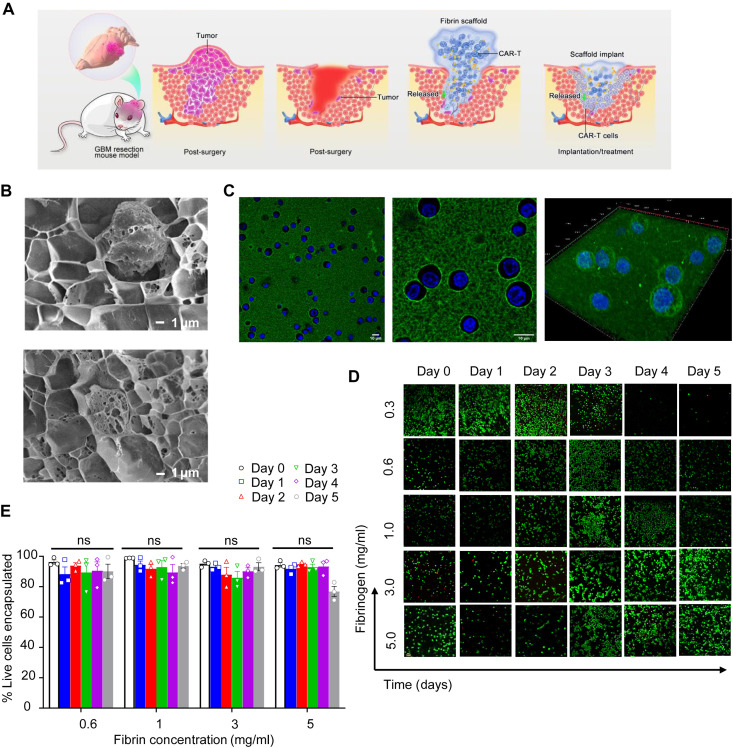
Fig. 1. Characterization of the fibrin gel delivery system for T cells.
(A) Schematic illustration of the procedure aimed at partially removing the GBM mass and delivering CAR-T cells via fibrin gel in situ formation. The fibrin gel in the inflamed surgery site creates the matrix, allowing the gradual release of CAR-T cells. (B) Cryo-SEM imaging of the CAR-T cells within the fibrin gel. Scale bars, 1 μm. (C) Confocal imaging of the CAR-T cells within the fibrin gel. CAR-T cells were labeled with green fluorescein, while the cell nuclei were stained with Hoechst 33254. High-resolution confocal imaging of cells encapsulated in fibrin gel is illustrated. Scale bars, 10 μm. (D) Representative confocal imaging showing live/dead CAR-T cells encapsulated within the fibrin network for 5 days. Live cells and dead cells were labeled with green fluorescein and red fluorescein, respectively. Scale bar, 1 μm. (E) Quantification of live CAR-T cells in the fibrin gel at different days and at different concentrations of fibrin as illustrated in (D); no difference [not significant (ns)] in viability for all concentrations.

