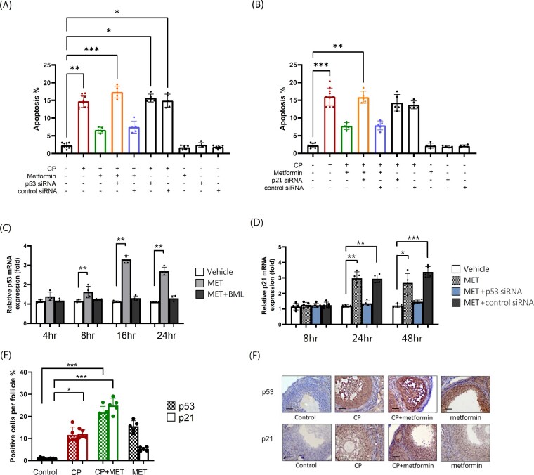
Figure 6.
Metformin exerted AMPK/p53/p21-mediated anti-apoptotic effect on cultured mouse granulosa cells. (A) Mouse granulosa cells were treated with p53 siRNA (25 nM) or control siRNA for 24 h prior CP (1ug/ml) or metformin (MET) (10 mM) treatment, after 72 h. Apoptotic cells were determined by flow cytometry with propidium iodide staining and quantified by the subG1 ratio. The experiments were repeated five times and therefore N = 5 per group. The apoptosis rate significantly increased in the CP-alone group (P = 0.0029) and tended to decrease after cotreating the cells with MET. The anti-apoptosis effect of MET was diminished after blocking the p53 activity with siRNA. (B) The experimental conditions were the same as A, except that p21 siRNA (25 nM) was applied instead of p53 siRNA. (C) Mouse granulosa cells were treated with AMPK inhibitor BML275(10 μM) for 30 min prior MET (10 mM) treatment. At indicated time periods, the expression of p53 mRNA was determined by qRT-PCR. The experiments were repeated four times and therefore N = 4 per group. Sequential measurement of p53 mRNA expression in culture granulosa cells significantly increased 8 h after the addition of MET (P = 0.0098). The expression of p53 mRNA was blocked with the addition of cell-permeable AMPK inhibitor BML. (D) The expression of p21 mRNA was determined by qRT-PCR and sequential measurement of p21 mRNA expression in cultured granulosa cells significantly increased 24 h after the addition of MET (P = 0.0095). The experiments were repeated five times and therefore N = 5 per group. (E) C57BL/6 mice were treated with CP-alone or in combination with MET or MET-alone. After 4 weeks of treatment, the ovaries were processed into paraffin sections for the p53 and p21 staining. Both p53 and p21 activity were generally low in normal untreated ovarian tissue but was elevated after CP treatment (p53: P = 0.6529; p21: P = 0.0452) as determined with IHC staining under microscopic examination at ×400 magnification. The coadministration of MET with CP increased p53 and p21 protein expression in the ovarian tissue even higher (P < 0.0004 in both). Statistical analyses were performed by nonparametric Kruskal–Wallis test with Dunn's post-hoc for multiple comparisons. *P < 0.05, **P < 0.01, ***P < 0.001. (F) Representative p53 and p21 IHC images of each group were shown. The scale bar is 50 μm.

