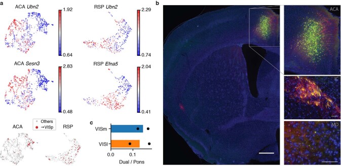
Extended Data Fig. 10. Validation of L5 ET + CC neurons.
a, UMAP of ACA (n = 1,131) and RSP (n = 516) L5 ET cells, coloured by gene body mCH of example genes. Ubn2 shows hypomethylation in the cluster enriching neurons projecting to the VISp in both ACA and RSP, whereas Sesn3 and Efna5 are hypomethylated in the cluster only in ACA or RSP, respectively. VISp-projecting cells are shown in red at the bottom. b, By injecting AAV-retro-Cre in the VISp and AAV-FLEX-GFP in the ACA, the axon terminals of ACA–VISp neurons were also observed in the internal capsule (IC) and mediodorsal nucleus of thalamus (MD). Scale bars, 500 μm (left) and 50 μm (right in IC and MD). c, The proportion of double-labelled neurons that project to both VISp and pons, out of neurons projecting to pons in medial and lateral visual cortex (VISm and VISl, respectively). n = 2 biological replicates are shown as individual points.

