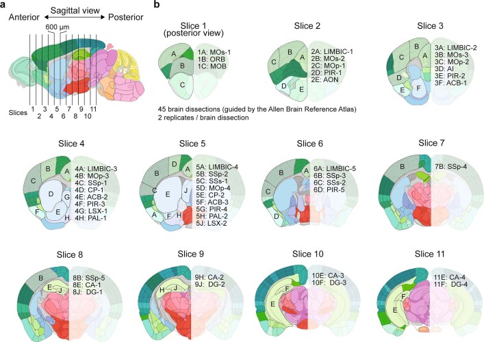
Extended Data Fig. 1. Anatomic maps of the 45 dissections in the adult mouse cerebrum.
a, Schematic of brain tissue dissection strategy. Mouse brains were cut into 600-μm-thick coronal slices. b, Brain regions dissected from each coronal slice are marked according to the Allen Brain Reference Atlas28. The frontal view of each slice from slices 1–11 is shown, with the dissected regions alphabetically labelled on the left, and the anatomic labelling listed on the right. A detailed list of the dissected regions and the full anatomic labelling can be found in Supplementary Table 1.

