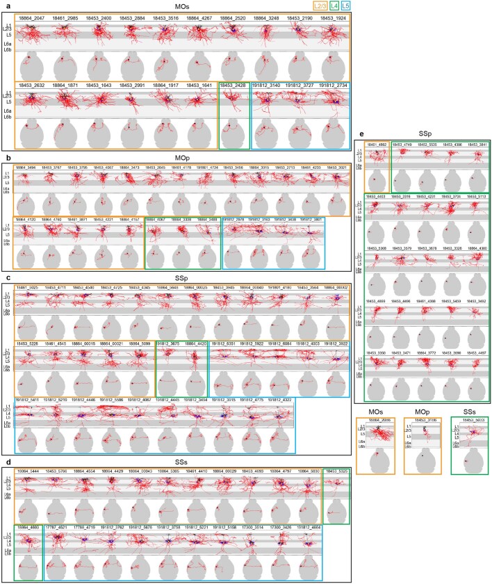
Extended Data Fig. 7. Local morphologies and long-range intracortical projections of cortical L2/3, L4 and L5 IT neurons.
a–d, Comparison of local morphologies (upper panels; apical dendrite in black, basal dendrite in blue, axon in red, soma as an orange dot) and intracortical projections (lower panels; axon in red, soma as a star) for MOs (a), MOp (b), SSp (c) and SSs (d) neurons. L2/3, L4 and L5 IT neurons are marked by orange, green and blue boxes, respectively. The L4-like neurons from MOp and MOs are located between L2/3 and L5 since L4 is not delineated in MOp or MOs in CCFv3. Neurons are ordered based on the depths from pial surface of their somas. Gray shadings mark generic layers; however, it should be noted that due to variation in layer thickness in different parts of the cortical areas, the generic layer marking does not necessarily correlate with each neuron’s precise soma location. The layer assignment of each neuron’s soma location was confirmed by visual inspection of each case. e, Reconstructed neurons without long-range axon projections outside of their soma areas. Vast majority of these neurons are SSp L4 IT. Overall, recent studies by scRNA-seq31, MERFISH66 and Patch-seq7 showed that transcriptomically defined cortical IT neuron types are organized by layer, but also exhibit a continuous spatial transition along the cortical depth. Here we arrange the L2/3, L4 and L5 IT neurons according to the depth of their soma from the pial surface, and find that within each region, across depths individual neurons exhibit highly variable long-range projection patterns. We identified 26 cells from SSp and 3 cells from SSs to be in L4. L4 cells have either no apical dendrites (i.e., spiny stellate cells) or a simple apical dendrite that does not branch in L1 (i.e., untufted or star pyramid cells), in contrast to the pyramidal L2/3 cells which have tufted or wide-branching apical dendrites in L167. L2/3 cells have local axons branching in L2/3 and downward into L5, whereas L4 cells have local axons mainly projecting up to L2/368. We also found 4 cells from MOp and MOs with these L4-like features – minimal apical dendrites and upward-projecting local axons, suggesting that these are the L4-like cells located in motor cortex69 that can also be identified transcriptomically70. Consistent with prior notion67, all but two SSp L4 cells have only local axons but no long-range projections. However, nearly all L4 cells in SSs, MOp and MOs do have axon projections outside of their local area, as we reported before33.

