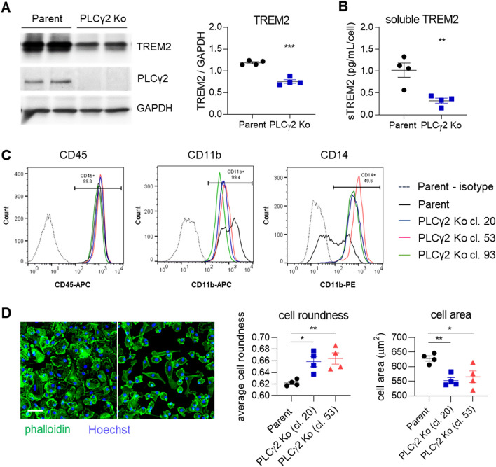
Figure 2.
PLCγ2 Ko leads to dysregulation of cell surface marker expression and morphological changes in iPSC-derived macrophages. (A) Western blot confirms lack of PLCγ2 protein in PLCγ2 Ko and shows reduction in TREM2 expression in PLCγ2 KO macrophages compared to Parent. n = 4. Full immunoblot images are presented in Supplementary Fig. 7. (B) Levels of soluble TREM2 detected in cell supernatant 8 days after plating are lower in PLCγ2 Ko cells compared to Parent. n = 4, (C) Macrophage surface markers CD11b, CD14, and CD45 were measured by flow cytometry, compared to relevant isotype IgG. Annotations indicate frequency of marker positivity in the Parent line. (D) Morphology of macrophage lines was determined by phalloidin staining and analysis of cell roundness and cell area. Scale bar 50 μm. n = 4, data shown represent mean ± SEM, One-way ANOVA followed by Bonferroni’s multiple comparison test. *p < 0.05, **p < 0.01.

