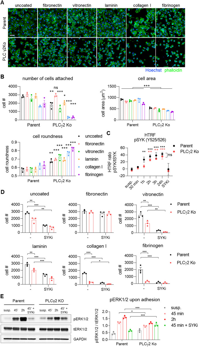
Figure 5.
Cell adhesion to different substrates is compromised in PLCγ2 Ko macrophages. (A) Representative images of Parent and PLCγ2 Ko cells attached to surfaces coated with the ECM molecules indicated. Scale bar 50 μm (B) Quantification of number of cells attached, cell area and cell roundness 3 h after plating on ECM molecules. n = 3; annotations compare Parent vs. PLCγ2 Ko for each coating. (C) Phosphorylation of SYK is increased after different time points upon adhesion, as opposed to cells kept in suspension (susp.) in Parent and PLCγ2 Ko line, n = 3; black annotations compare Parent adhered vs. suspension, red annotations compare PLCγ2 Ko adhered vs. suspension (D) Effect of SYK inhibition using BIIB-057 (SYKi) on cell adhesion, demonstrating SYK-dependency of adherence to uncoated plates, vitronectin, collagen I, laminin and fibrinogen, but not to fibronectin. n = 3 (E) Representative Western blot showing that phosphorylation of ERK1/2 upon adhesion is decreased in PLCγ2 Ko cells. Full immunoblot images are presented in Supplementary Fig. 11. n = 3, data shown represent mean ± SEM, two-way ANOVA followed by Bonferroni’s multiple comparison test. *p < 0.05, **p < 0.01, ***p < 0.001.

