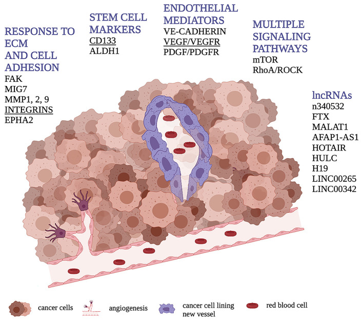
Figure 1.
Schematic representation of VM through cancer cells (in purple) forming a vessel containing red blood cells. Figure shows the main molecular pathways involved in the VM process in human osteosarcoma highlighting, in underlined bold, those found to be related with VM presence or tubular/vessel-like formation in vitro in dog.

