Figure 3.
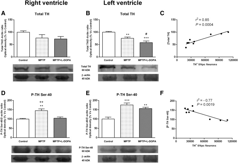
Effects of MPTP and L-DOPA treatment on total TH and phospho-TH at Ser-40 expression in the monkey heart. (A, B) Total TH/β-actin ratio (optical density, % vs control) in controls, MPTP-treated monkeys and in the MPTP + L-DOPA group. Total TH expression was significantly decreased in the LV in MPTP-treated monkeys and in the MPTP + L-DOPA group versus control group and in the MPTP + L-DOPA group versus MPTP-treated animals. (C) Strong correlation between the number of TH + neurons in the SNpc and total TH levels in the heart tissue (r2 = 0.85, P = 0.0004). (D, E) P-TH at Ser-40/β-actin ratio (optical density, % vs control) in controls, MPTP-treated monkeys and in the MPTP + L-DOPA group. TH phosphorylation at Ser-40 was significantly increased in the RV with respect to the other two groups. Besides, in the LV, we found an important increase of phospho-TH at Ser-40 in both groups (MPTP or MPTP + L-DOPA treatment) with respect to controls. (F) Strong correlation between phospho-TH and TH+ neurons in the SNpc (r2 = 0.77, P = 0.0019). Data were compared by one-way ANOVA (total TH: F(2,9) = 2.063, P = 0.1831, RV; F(2,9) = 17.87, P = 0.0007, LV. P-TH: F(2,9) = 12.77, P = 0.0024, RV; F(2,9) = 17.43, P = 0.0008, LV) followed by Newman–Keuls post-hoc test. Values are represented as mean ± standard deviation. **P < 0.01, ***P < 0.001 versus control group; #P < 0.05 versus MPTP-treated animals; ++P < 0.01 versus MPTP + L-DOPA. TH tyrosine hydroxylase, MPTP 1-methyl-4-phenyl-1,2,3,6-tetrahydropyridine, L-DOPA levodopa, P phospho. (n = 4 animals/group and 2 technical replicates/animal).

