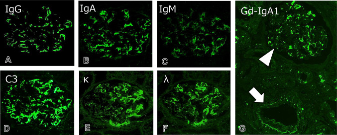
Fig. 2.
Immunofluorescence findings. A–D Immunofluorescence study shows the granular mesangial deposition of IgA, IgG, IgM, and complement C3 in glomeruli (original magnification, × 600). E, F Both κ and λ light chains are noted as a mesangial granular pattern (original magnification, × 600). G The deposition of galactose-deficient IgA1 (Gd-IgA1) is evident in mesangial areas in glomeruli (arrowhead), compatible with IgAV. A small arteriole without necrotizing arteritis is negative for Gd-IgA1 (arrow) (original magnification, × 200)

