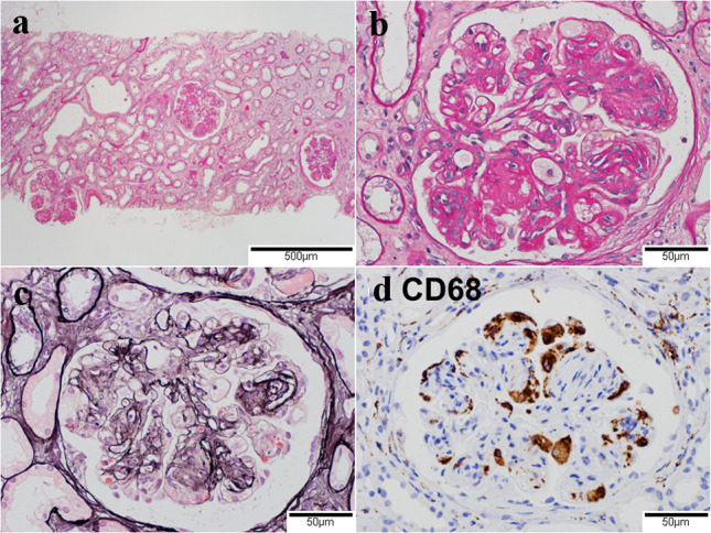
Fig. 4.

Light microscopic findings in the glomerulus. a Nodular lesions in glomeruli at low magnification. Mild infiltration of inflammatory cells in the renal interstitium and tubular atrophy. (Periodic acid-Schiff stain, original magnification ×40). b Lobular accentuation of the glomerular capillary tufts with marked mesangial expansion and capsular adhesion. Infiltration of foam cells in glomerular capillaries. (Periodic acid-Schiff stain, original magnification ×200). c Expansion of subendothelial spaces and double contours in the glomerular capillary walls. (Periodic acid-methenamin-silver stain, original magnification ×200). d CD68 positive infiltrating cells in the glomerular capillaries. (Immunoenzyme stain, original magnification ×200)
