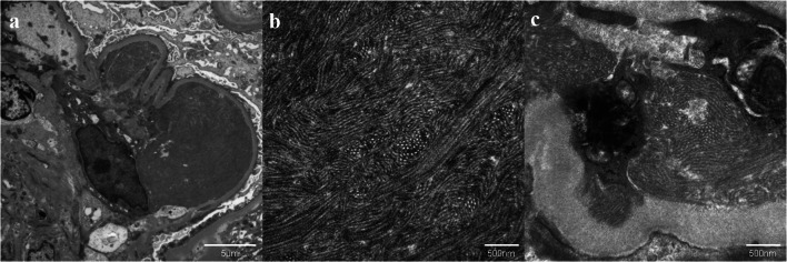
Fig. 6.
Electron microscopic findings in the glomerulus. a Large amounts of organized deposits in the subendothelial spaces (original magnification ×3000). b Higher magnified image of these deposits. Randomly arranged microtubular structures with 40–50 nm diameter. (original magnification ×20,000). c Similar microtubular structures in another subendothelial space. (Original magnification ×20,000)

