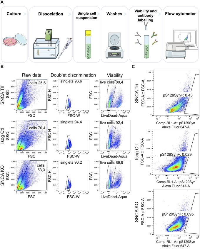
Figure 6.
pS129Syn detection in SNCA Tri hMOs. (A) Flow cytometry sample processing schematic. Three organoids per genotype were incubated into enzymatic solution at 37°C, then manually dissociated with a pipet. Single-cell suspensions were obtained after the filtration of dissociated tissues. The single-cell suspensions were labelled for cell viability and antibodies against internal and external epitopes, before signals were read by the flow cytometer. (B) Gating strategy for the removal of cellular debris, doublet discrimination and cell viability. (C) Proportion of total cells carrying pS129Syn in single-cell dissociated hMOs (3 hMOs pooled per line) measured by flow cytometry.

