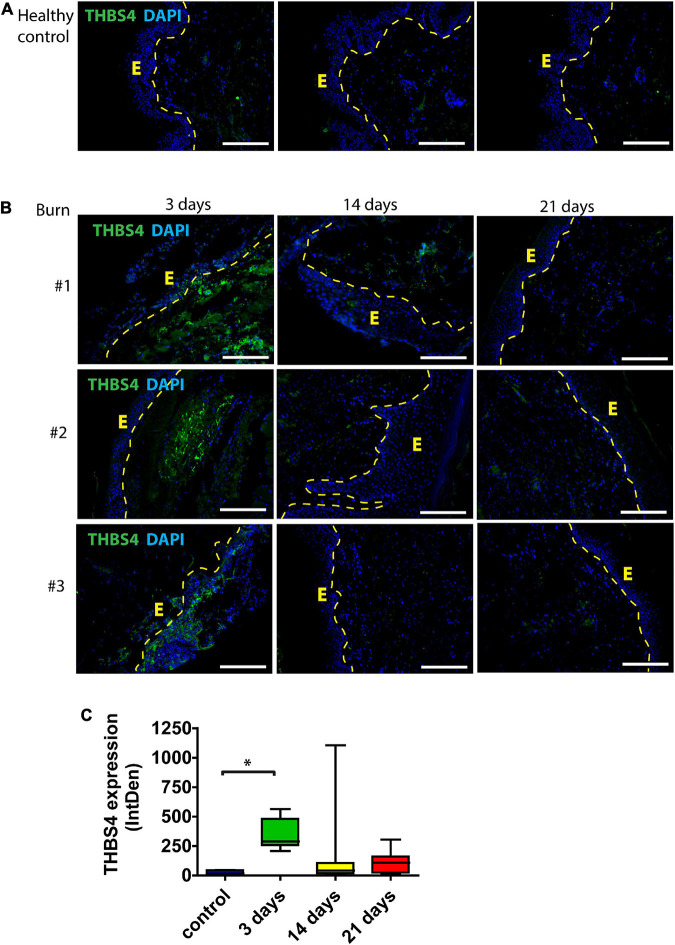
FIGURE 1.
THBS4 expression in human skin following burn injury. (A) Normal skin from healthy controls; (B) skin from burn injury patients. Biopsy samples were collected from the study area at 3, 14, and 21 days after the wound excision. E—epidermis. 3 representative samples in each group are shown. Scale bar is 200 μm. (C) Relative quantification of THBS4 expression by mean integrated density of the fluorescence signal. The plot depicts the distribution of 10 samples, * indicates a statistically significant (P < 0.05) difference.

