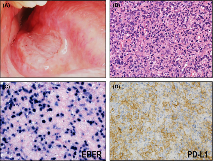
FIGURE 3.

Macroscopic, histological, and immunohistochemical features of EBV‐positive mucocutaneous ulcer (EBVMCU). (A) EBVMCU macroscopically shows a sharply circumscribed mucosal ulcer. (B) Histologically, EBVMCU shows infiltration of intermediate to large atypical lymphoid cells with polymorphous background (HE ×400). (C) EBER is detected in the large atypical lymphoid cells (×400). (D) Most of the cases are negative for PD‐L1 on tumor cells (×400)
