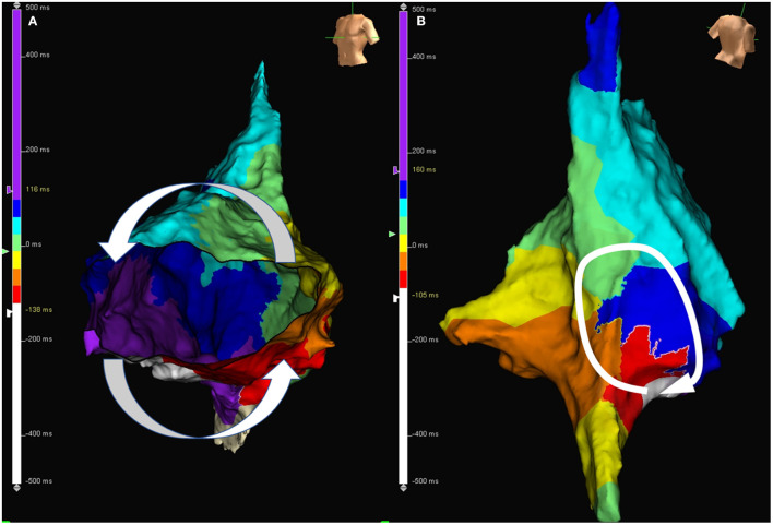
Figure 2.
(A) Activation map during AFL in a patient with typical AFL. The activation wave front travels through cavotricuspid isthmus and goes around the tricuspid annulus. (B) Activation map during atypical atrial flutter in a patient with atypical AFL. The activation wave front goes around the surgical scar located at the right posterior free wall. AFL, atrial flutter.

