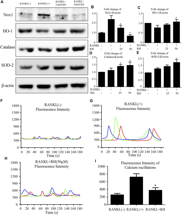
FIGURE 5.
RH suppressed RANKL-induced ROS activity and Ca2+ oscillation. (A) Representative Western blot images of the Nox1, catalase, HO-1, and SOD-2 expression with the treatment of RH, the Nox1 expression was significantly suppressed by RH, and the antioxidant enzymes of HO-1, catalase and SOD-2 were enhanced. BMMs were stimulated with RANKL (50 ng/mL) with RH at the concentration of 25 and 50 μM or PBS for 2 days before collecting protein. (B–E) Quantification of the ratios of band intensity of Nox1, HO-1, catalase, and SOD-2 relative to β-actin (n = 3 per group). *p < 0.05, **p < 0.01 comparison with the RANKL-induced positive control group. (F–H) Representative images of fluorescence intensity waves of Ca2+ oscillation in negative group, RANKL stimulated positive group and RH (50 μM) treated group. There were three colours indicating different cells in each group. (I) Quantification of fluorescence intensity change of Ca2+ oscillation in each group. *p < 0.05, **p < 0.01 relative to RANKL-induced positive control group.

