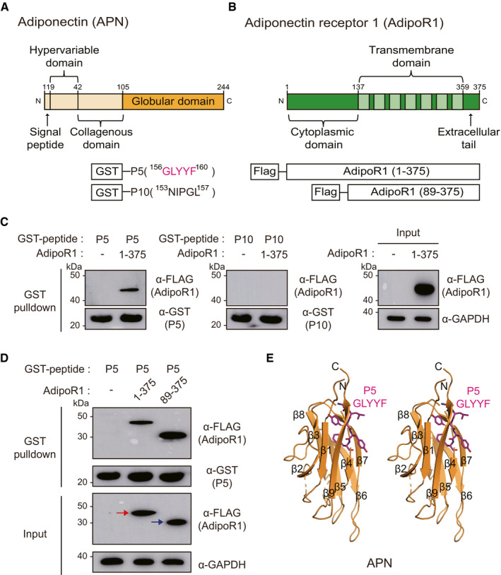
Figure 1. Direct molecular interaction between the P5 and AdipoR1.

- Domain architectures of APN and the GST‐fused 5‐mer peptides (GLYYF; GST‐P5, or NIPGL; GST‐P10).
- Domain architectures of AdipoR1 and Flag‐fused full‐length AdipoR1 (residue 1–375) or N‐terminal‐truncated AdipoR1 (residues 89–375). Each construct in the schematic diagram was used for the pulldown experiment.
- GST pulldown of transiently expressed AdipoR1 (residues 1–375) with immobilized GST‐P5 or GST‐P10 fusion proteins. Pulldown fractions were subjected to Western blotting using Flag and GST antibodies.
- GST pulldown of AdipoR1 (residues 1–375) or AdipoR1 (residues 89–375) with GST‐P5. The red or blue arrow indicates correct sized bands of AdipoR1 (residues 1–375) or AdipoR1 (residues 89–375), respectively.
- The position of P5 in APN. A stereo ribbon diagram of APN (PDB ID: 4DOU) is drawn, and P5 is shown in magenta sticks.
Source data are available online for this figure.
