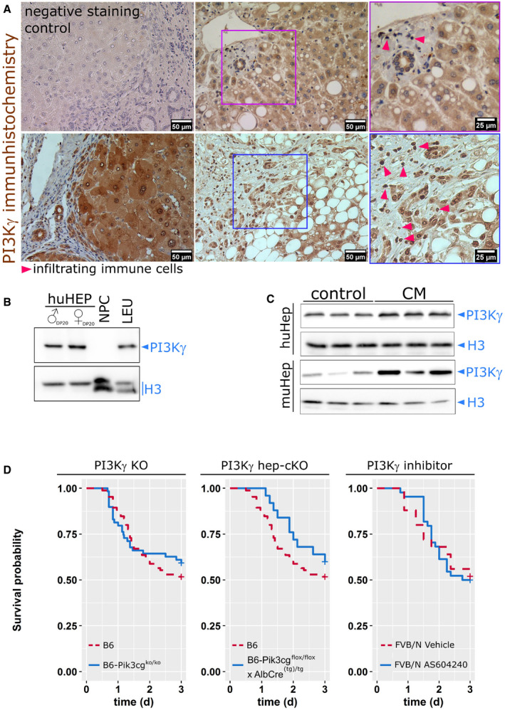
Figure 1. Cell‐type‐specific expression pattern of PI3Kγ in the liver, induction, and function under inflammatory conditions.

- PI3Kγ is expressed by human hepatocytes and infiltrating immune cells in biopsies from patients with minimal to mild inflammatory activity. Triangles point to immune cells (clusters), including some neutrophils known to express PI3Kγ highly. In the negative control, the primary antibody was replaced by an equal volume buffer. The number of included patients, gender, and diagnosis are summarized in Appendix Table S1.
- PI3Kγ expression in human primary hepatocytes from 20 male (♂) or female (♀) donor pools (HEP, DP20), but not non‐parenchymal cells (NPC). Isolated human leukocytes from healthy volunteers (LEU) served as positive controls. The characterization of the donor pools is summarized in Appendix Table S2.
- Primary murine hepatocytes and HepG2 cells expressed PI3Kγ under basal conditions; expression was increased at 24 h after stimulation with LPS, IFN‐γ, IL‐1β, and TNF‐α (CM).
- Survival of WT, PI3Kγ null (left) and liver‐specific PI3Kγ knockout mice (PI3Kγ floxtg/tg × AlbCre(tg)/tg, middle) or systemic application of the PI3Kγ inhibitor, AS605240 (right) in a model of polymicrobial sepsis induced by peritoneal contamination with a human stool suspension. Animal numbers: B6: 86 (%male: 52, %female: 48), B6‐Pi3kcgko/ko: 59 (%male: 51, %female 49), B6‐Pi3kcgflox/flox × AlbCre(tg)/tg: 25 (%male: 68, %female 32), FVB/N Vehicle: 25 (%male: 44, %female 56), FVB/N AS: 43 (%male: 44, %female 56).
