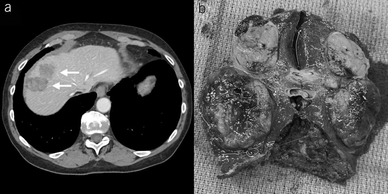
Figure 1.

(a) Two masses (arrows) measuring 3.2 by 2.4 cm and 2.3 by 2.1 cm with a heterogeneous signal and washout of contrast. (b) Gross liver specimen including segments 4 and 8 measuring 9×9× 4 cm. A reddish-brown soft round mass measuring 3 × 3 × 2.5 cm and yellowish-gray oval firm mass measuring 2×2×1.5cm are visualized. An additional nodule measuring 1×0.8×30.8cm was identified by dissection (not visualized on gross specimen).
