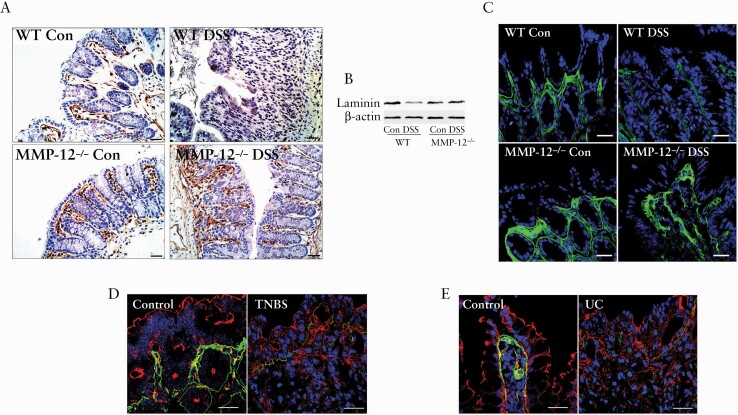
Figure 6.
Immunolocalisation of laminin in intestinal inflammation. A: In immunohistochemical examination, polyclonal laminin antibody detected the basement membrane [brown] below the epithelial lining of control colon in WT and MMP-12-/- mice. The laminin staining at the basement membrane was found to be completely disappeared in WT DSS colon but not in MMP-12-/- DSS mice colon. Nuclei are counterstained with haematoxylin. Black bar = 30 μ m. B: In western blotting, laminin protein expression was significantly reduced in WT DSS colon and not in MMP-12-/- DSS colon. Representation of three blots in each group. C: The laminin α1-specific staining [green] in the basement membrane was found to be markedly lost in WT DSS colitis but not in MMP-12-/- DSS colitis. The laminin α1 expression [green] was found to be lost in murine TNBS colitis [D] and in UC [E], compared with respective controls. Actin is stained in red and nuclei are stained in blue. White bar: 50 μ m. Representation of several microscopic areas, from three or more samples in each group. DSS, dextran sodium sulphate; WT, wild-type; TNBS, trinitrobenzene sulphonic acid; UC, ulcerative colitis.

