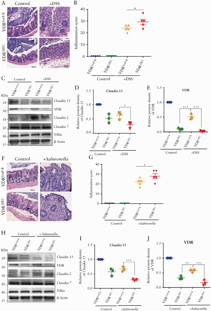
Figure 8.
Reduced Claudin-15 expression due to deletion of VDR in the intestinal epithelium in the experimental colitis. [A] Representative images of H&E of mice with or without DSS treatment. [B] Increased inflammation in VDRΔIEC mice was noted as compared with VDRloxP-B. [C] Representative images of western blot of Claudin-15, Claudin-2, Claudin-7, and VDR after DSS treatment. Densitometric analysis of Claudin -15 [D] and VDR [E] in VDRΔIEC mousee colon in DSS colitis. [F] Representative images of H&E of mouse caecum 4 days post Salmonella infection. [G] Increased inflammation in VDRΔIEC mice was noted as compared with VDRloxP-B after Salmonella infection. [H] Representative images of western blot of Claudin-15, Claudin-2, Claudin-7, and VDR following Salmonella treatment. Densitometric analysis of Claudin -15 [I] and VDR [J] in the colon of VDRΔIEC mice in Salmonella-induced colitis. Scale bar = 50 µm, n = 3–6. Data were analysed by unpaired t test or one-way ANOVA, *p ≤ 0.05, **p ≤ 0.01, and ****p ≤ 0.001. VDR, vitamin D receptor; H&E, haematoxylin and eosin; ANOVA, analysis of variance.

