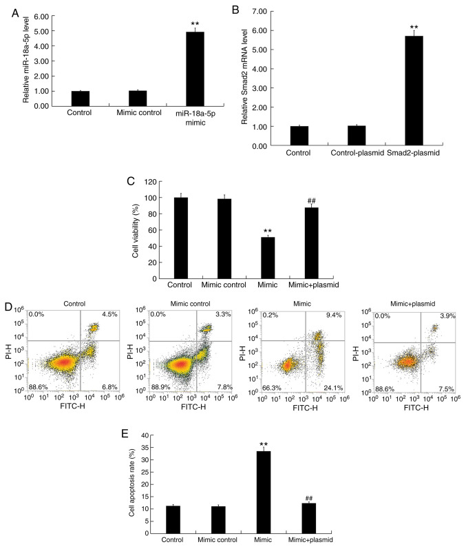
Figure 5.
Effect of miR-18a-5p mimic on hHSFs. (A) hHSFs were transfected with mimic control or miR-18a-5p mimic for 48 h, and then RT-qPCR was used to detect the miR-18a-5p level. (B) hHSFs were transfected with control-plasmid or Smad2-plasmid for 48 h, and then RT-qPCR was used to detect Smad2 mRNA level. (C) hHSFs were transfected with mimic control, miR-18a-5p mimic or miR-18a-5p mimic + Smad2-plasmid for 48 h, and then MTT was used to detect cell viability. (D) Flow cytometry graphs and (E) cell apoptosis rate in hHSFs transfected with mimic control, miR-18a-5p mimic or miR-18a-5p mimic + Smad2-plasmid for 48 h. Data are presented as the mean ± standard deviation. **P<0.01 vs. control group; ##P<0.01 vs. mimic group. miR, microRNA; hHSFs, human hypertrophic scar fibroblasts; RT-qPCR, reverse transcription-quantitative polymerase chain reaction.

