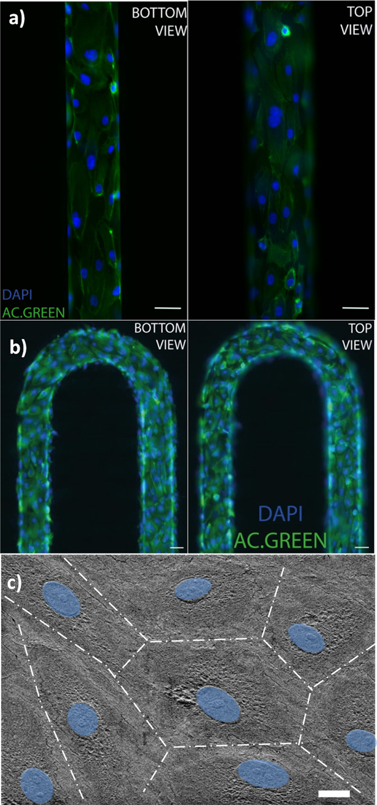
Figure 2.

Fluorescence images of the HUVEC adhesion with actin (green) and nucleus (blue) covering the surface of the microchannels obtained from (a) a photolithography master mold and (b) a 3D-printed master mold (scale bar = 100 μm). (c) SEM images of HUVECs on a microfluidic device (nucleus colored in blue) (scale bar = 10 μm).
