
. 2021 May 27;8(1):G87–G136. doi: 10.1530/ERP-20-0034
 This work is licensed under a
 Table 8.
Characteristics that identify unfavourable outcome to MV repair surgery
| View | Measure or image | Explanatory note | Image |
|---|---|---|---|
| PLAX |
Image 1
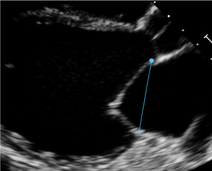
Annular diameter |
Measure A-P annular diameter in systole |
|
| All views |
Image 2
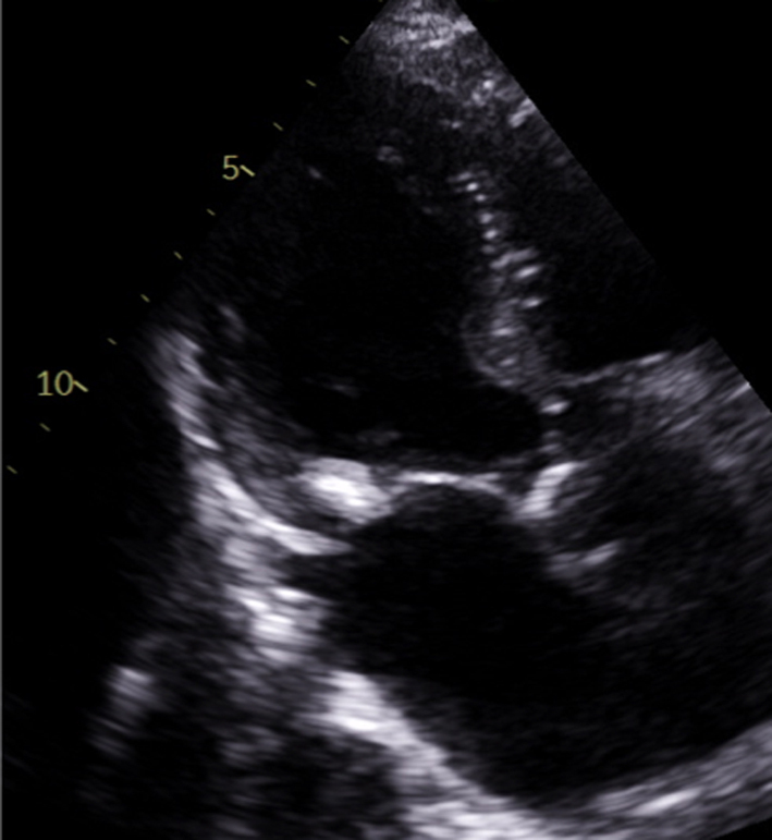
Extent of calcification |
Describe the location and extent of calcification in the annulus, leaflets and subvalvar apparatus |
|
| PLAX |
Image 3
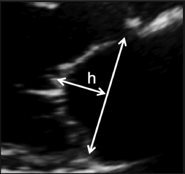
Coaptations height |
Zoom on the mitral valve in the parasternal long-axis view. Freeze the image and scroll through to mid-systole. Draw a line between the anterior and posterior annular points. Measure the coaptation height perpendicular to the plane of the annular line. |
|
| PLAX |
Image 4
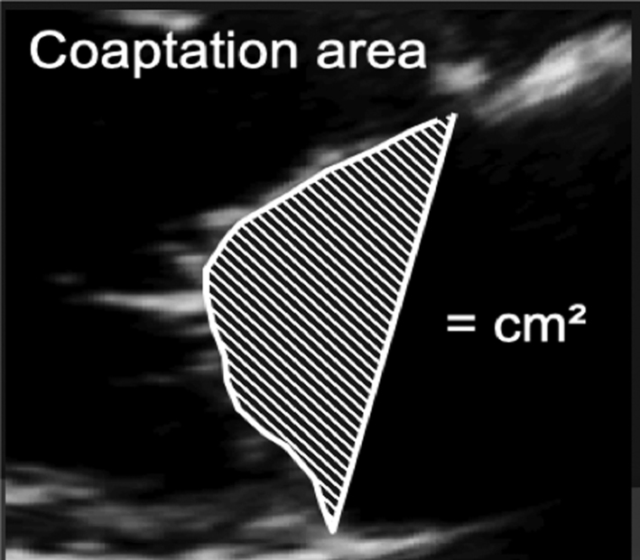
Coaptation area |
Once coaptation height has been measured, the area between the annular plane and the atrial surface of the leaflets can be measured. |
|
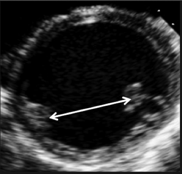
| PSAX – PM |
Image 5
Inter-papillary distance |
Freeze the image and scroll to end-systole. Measure the inter-papillary distance. |
|
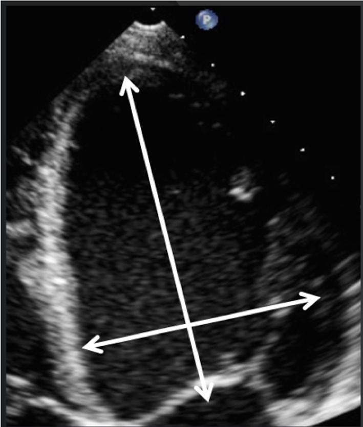
| A4C |
Image 6
Systolic sphericity index |
Adjust the depth of the image to focus on the LV. Freeze the image and scroll to peak systole. Measure the diameter and longitudinal dimension at the longest/widest. Divide the basal diameter by the longitudinal dimension, a value >0.7 Indicates adverse LV remodelling. |
|