
Figure 2 |.
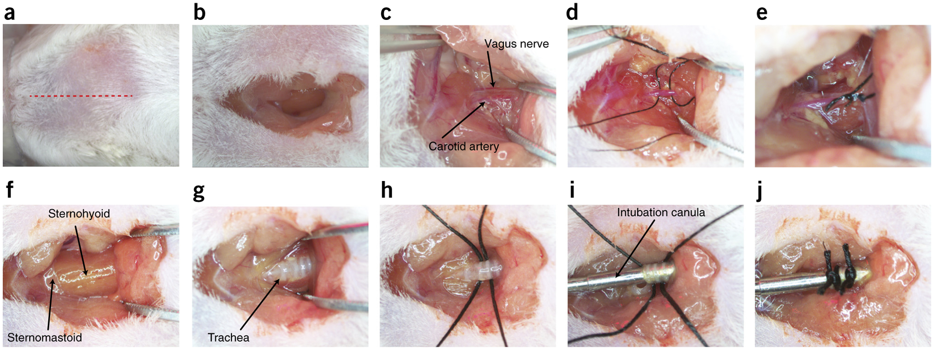
Surgical preparation of carotid to artery ligation and tracheotomy. (a) Red dotted line indicates location of superficial cut along the midline of the neck. (b) Excavation of the superficial adipose tissue to expose the sternohyoid muscles. (c) Blunt dissection of the carotid artery and vagus nerve. (d) 6-0 sutures run underneath the carotid artery, avoiding the vagus nerve. (e) Unilateral tied sutures of the carotid artery. (f) Location of sternohyoid and sternomastoid muscles. (g) Separation of the sternohyoid muscle to expose the trachea. (h) 4-0 sutures run under the trachea for future use. (i) Tracheal cannula inserted into the trachea. (j) Tracheal cannula sutured into place for attaching to a ventilator. Local ethics committees approved the procedures shown here. This figure illustrates Steps 5–14 of the PROCEDURE.