
Figure 7 |.
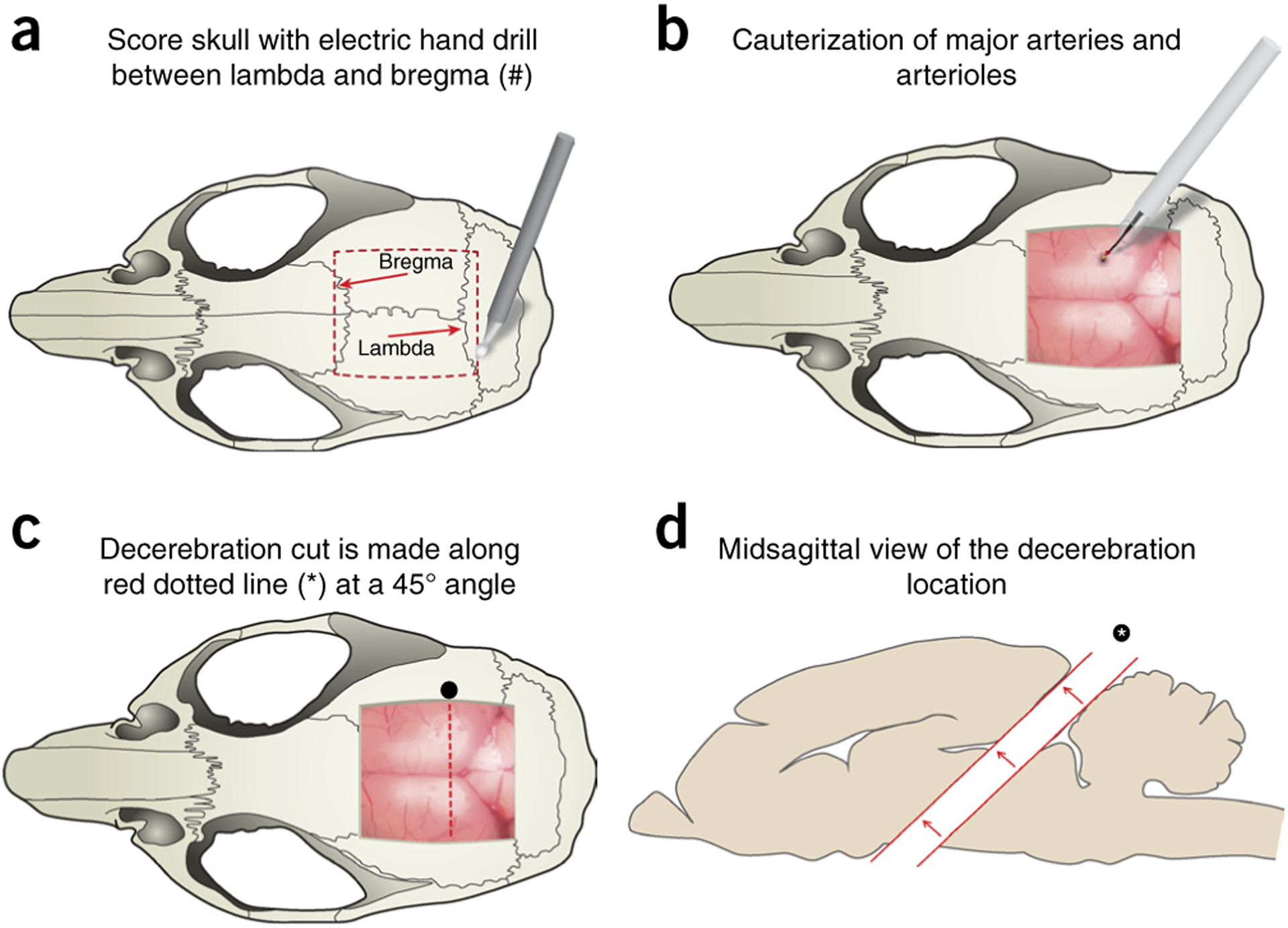
Craniotomy and decerebration cut location. (a) Craniotomy and brain exposure are performed by first scoring a square outline (red dashed box) between the lambda and the bregma using an electric handheld drill. The mouse rongeur tool is used to remove the skull as one piece by lifting the scored area, exposing the brain and meninges. (b) Superior sagittal sinus vein and other major vasculature are cauterized using hand cauterization tool (Acu-Tip Portable or Bovie Change-A-Tip) at the most caudal end to reduce bleeding. (c) Schematic representation of the removal of the cerebral cortex (decerebration) and underlying structures. Decerebration is performed with a no. 10 round blade, cutting through the brain at a 45° angle; cut is represented by an asterisk and the red dotted line. (d) Rostral portion of brain is carefully removed from the cut (asterisk) using a microspatula and without damage to the remaining caudal section. In the absence of the rostral brain, the remaining cavity is filled with a hemostatic sponge and/or Surgicel. This figure illustrates Steps 20–27 of the PROCEDURE. Local ethics committees approved all procedures shown here.