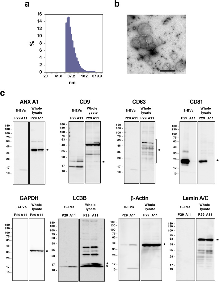
Fig. 5.
Characterization of S-EVs isolated from the conditioned medium of A11 cells. (a) A percent bar plot showing the distribution of particle diameter of A11 S-EVs. (b) Negative-stain TEM of A11 S-EVs. Bar: 500 nm. (c) Western blot analyses of various proteins in S-EVs isolated from the conditioned medium of P29 and A11 cells. P29 and A11 S-EVs (3 μg proteins) and whole cell lysates of P29 and A11 cells (30 μg proteins) were loaded on the same gel. Asterisks indicate the position of individual protein. In the LC3B immunoblot, the upper and lower asterisks indicate LC3-I and LC3-II, respectively. Uncropped Western blots images are shown in Fig. S7

