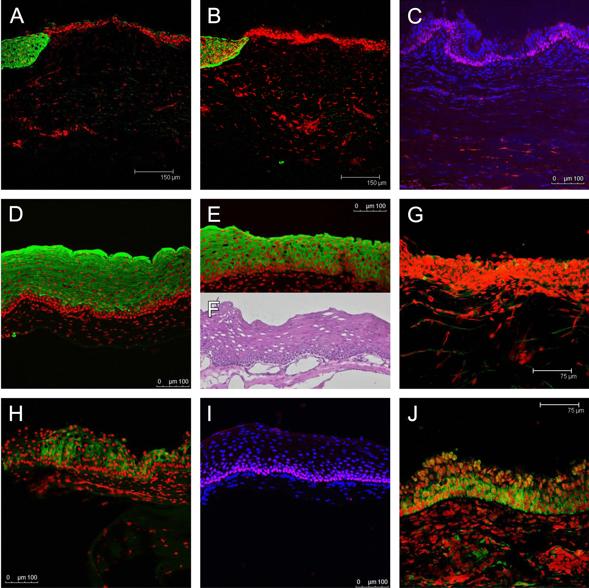
Fig. 4.

Immunoconfocal microscopy for keratin 12 (A, green; nuclei counterstained with PI), 8 (B, green), and p63 (C, red; nuclei counterstained with DAPI) in a corneal button removed half a year after COMET, and keratin 4 (D, green), 13 (E, green), 8 (G, green; no signal seen), 3 (H, green), p63 (I, red) and p75NTR (J, green) in a limbal biopsy taken 2 years after COMET. Keratin 12 (A) and keratin 8 (B) were both positive in the upper part (left side) of the corneal button, suggesting the presence of corneal epithelium in this area. While the majority of the specimen was keratin 12 and keratin 8-negative oral mucosal epithelium, p63 signal was universally expressed in the basal epithelium (C). Biopsy from the lower limbus 2 years after COMET showed stratified epithelium with over 10 layers of cells in some areas (F). The basal epithelial cells were small, compact, and with a high N/C ratio. The epithelium was positive for keratin 4 (D), 13 (E), and 3 (H) in the suprabasal layer but uniformly negative in the basal layer. Negative keratin 8 staining confirmed the origin from the oral mucosa (G). Staining for p63 (I) and p75NTR (J) was both uniformly positive in the basal layer
