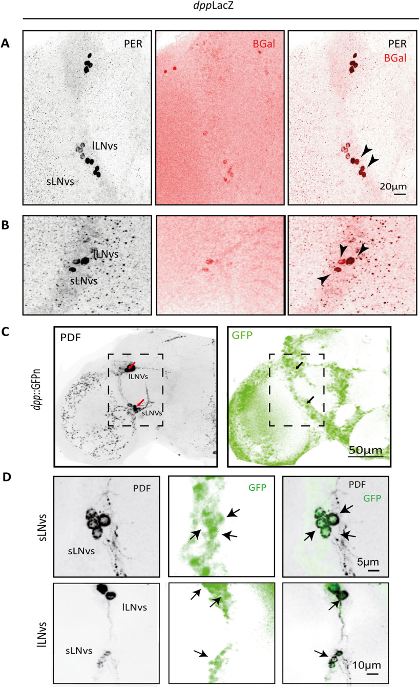
Figure 2.
DPP is expressed in the LNv cluster. A, B, Representative confocal images of two hemibrains showing the detection of DPP with the reporter line dppLacZ. The sLNvs and lLNvs were identified by PER immunoreactivity (in gray) and DPP through β-Galactosidase staining (β-Gal, in red). Black arrowheads indicate localization of the two signals in the same groups of cells. C, D, Representative confocal images of a different reporter line, dpp::GFPn; the PDF signal (left, gray) and GFP signal (right, green) are shown. D, Higher magnification of the sLNv and lLNv cluster from a different brain. Arrows highlight DPP reporter accumulation in the LNvs.

