FIGURE 1.
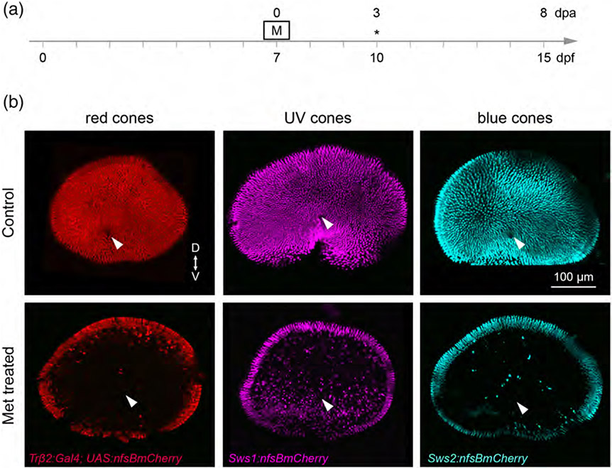
Selective ablation of specific cone populations in larval zebrafish. (a) Timeline demonstrates timing of Met (M) treatment. Asterisks denotes the age at which larvae were fixed for analysis. (b) En face views of wholemount, fixed retinas from 10 dpf (3 dpa) Met-treated and control fish. Met was applied for 1 hr at 7 dpf. Specific cone populations were targeted for ablation by selective expression of nfsB. Tg(trβ2:G4VP16; UAS:nfsBmCherry) fish were used to ablate red cones, Tg(sws1:nfsBmCherry) fish were used to ablate UV cones, and the Tg(sws2:nfsBmCherry) line was used to ablate blue cones. Arrowheads denote the optic nerve head. (“D” dorsal, “V” ventral)

