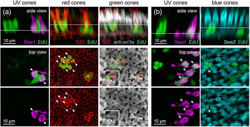
FIGURE 5.
Example of identification of regenerated cone types after UV cone ablation. Demonstration of how different cone types were distinguished in the population of regenerated photoreceptors after UV cone ablation. (a) Identification of regenerated UV, red, and green cones after UV cone ablation in 15 dpf (8 dpa) Tg(sws1:nfsBmCherry; trβ2:MYFP) double transgenic fish, with arrestin3a immunostaining and EdU labeling. Panels shown are images taken from the same region of tissue. (b) Identification of regenerated UV and blue cones after UV cone ablation in 15 dpf (8 dpa) Tg(sws1:nfsBmCherry; sws2:GFP) double transgenic fish with EdU labeling. Panels shown are images taken from the same region of tissue. In (a) and (b), side views are orthogonal rotations of the ONL from UV cone-ablated retinas. Top views show the nuclei located at the level of the line indicated in the side view. In order to visualize EdU-positive UV cones, a different plane of section was visualized because UV cone cell bodies reside in a lower plane compared to other cone types. Arrowheads point to EdU-positive nuclei

