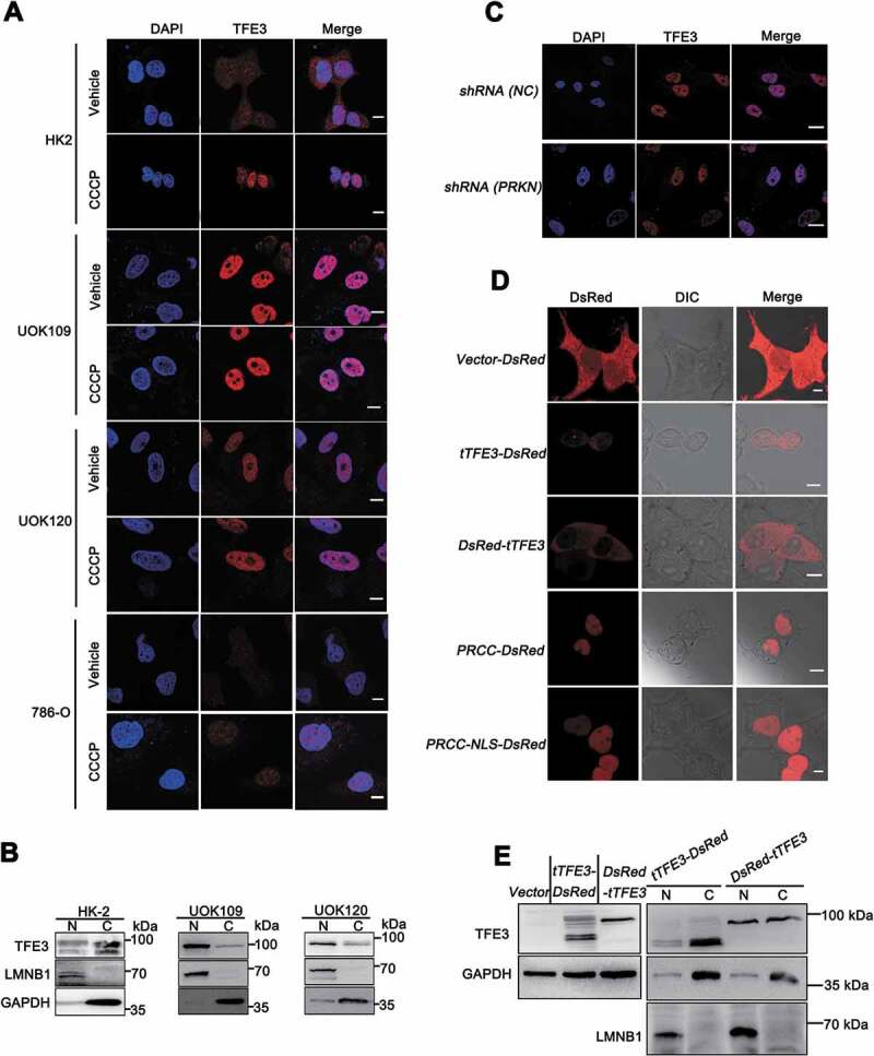
Figure 4.

Nuclear translocation of PRCC-TFE3 escapes from the regulation of PRKN-dependent mitophagy. (A) HK-2, UOK109, UOK120, or 786-O cells were treated with vehicle (DMSO) or CCCP (10 μM) for 6 h before immobilization and staining with a fluorescent anti-TFE3 antibody (red), followed by immunofluorescence photomicrographic analysis. Scale bars: 10 μm. (B) HK-2, UOK109 and UOK120 cells were fractionated and TFE3 was detected by western blot in nuclear fraction and cytoplasm fraction. N, nuclear fraction; C, cytoplasm fraction. (C) UOK120 cells were infected with Lenti.shRNA (NC) or Lenti.shRNA (PRKN) before immobilization and staining with a fluorescent anti-TFE3 antibody (red), followed by immunofluorescence photomicrographic analysis. Scale bars: 20 μm. (D) HEK293T cells were transiently transfected with constructs expressing PRCC (exon1)-RFP, PRCC (NLS)-RFP, RFP-tTFE3 (TFE3 fragment of PRCC-TFE3), or tTFE3-RFP, followed by visualization of RFP distribution using live-cell imaging microscopy. Scale bars: 10 μm. (E) RFP-tTFE3- and tTFE3-RFP-transfected cells as in (D) were fractionated and tTFE3 was detected by western blot in nuclear fraction and cytoplasm fraction. N, nuclear fraction; C, cytoplasm fraction
