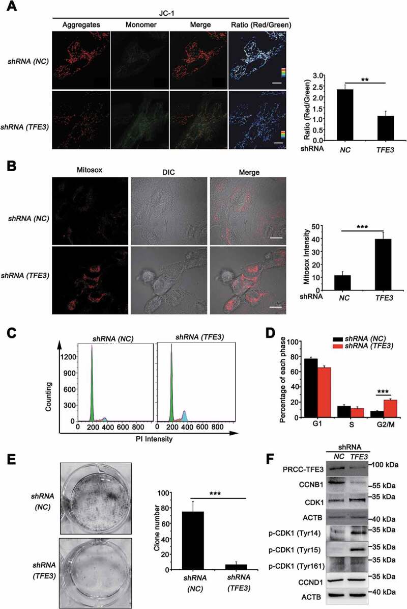
Figure 8.

PRCC-TFE3 fusions are involved in cell proliferation through regulating mitochondrial ROS formation. UOK120 cells were infected with Lenti.shRNA (NC) or Lenti.shRNA (TFE3) followed by staining with a JC-1 probe (A) or Mito-Sox (B) for 30 min. JC-1 or Mito-Sox intensity was measured with live-cell imaging microscopy. Data are presented as mean ± s.e.m from 3 independent experiments (10 cells/group/experiment in A and B). *p < 0.05, **p < 0.01, ***p < 0.001 (two-tailed t-test), Scale bars: 20 μm (in A) and 30 μm (in B). (C) UOK120 cells were infected with Lenti.shRNA (NC) or Lenti.shRNA (TFE3) and DNA content was determined by propidium iodide (PI) staining using flow cytometry. (D) Percentage of cells in each phase was quantified. Data are presented as mean ± s.e.m from 3 independent experiments. *p < 0.05, **p < 0.01, ***p < 0.001 (two-tailed t-test). (E) UOK120 cells were infected with Lenti.shRNA (NC) or Lenti.shRNA (TFE3), and then formation of colonies was measured after 7 d incubation. Data are presented as mean ± s.e.m from 3 independent experiments. *p < 0.05, **p < 0.01, ***p < 0.001 (two-tailed t-test). (F) UOK120 cells were infected with Lenti.shRNA (NC), Lenti.shRNA (TFE3) and then PRCC-TFE3, CCNB1, CCND1, CDK1, p-CDK1 (Tyr14), p-CDK1 (Tyr15) and p-CDK1 (Tyr161) were examined by western blot
