
Figure 4.
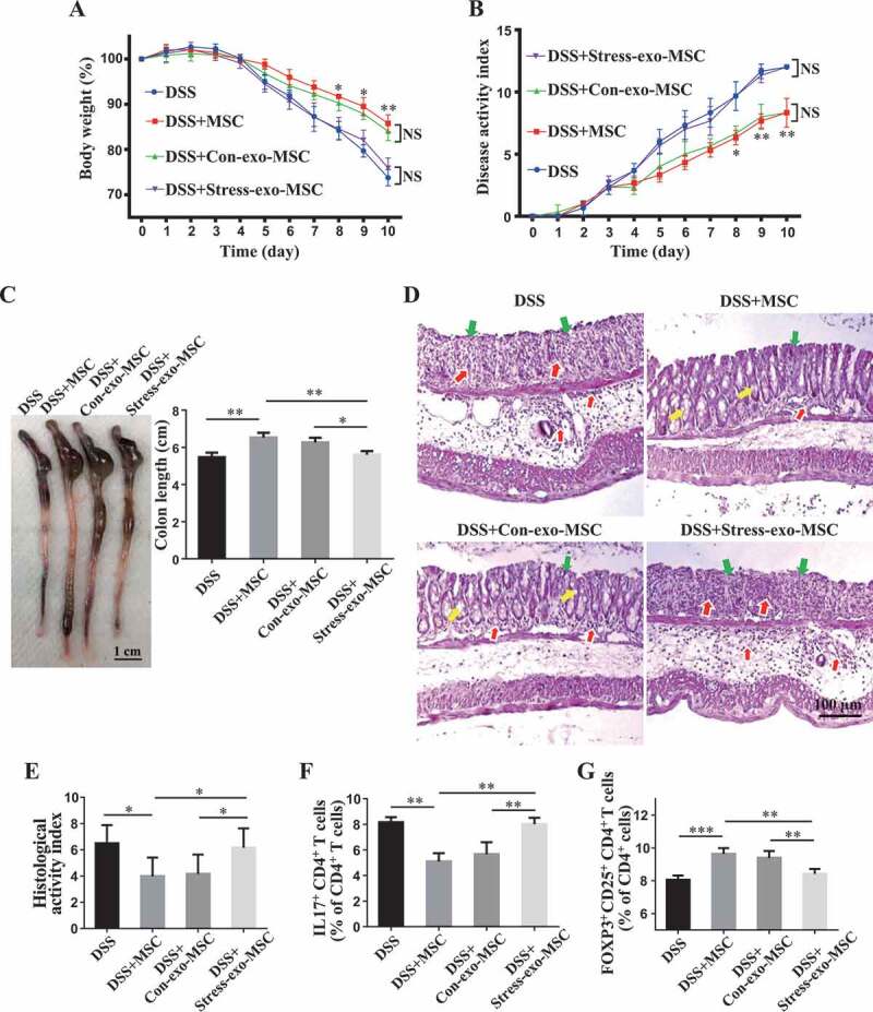
Circulating exosomes altered by restraint stress inhibit therapeutic effects of MSCs in DSS-induced colitis. (A-D) At 3 days after DSS treatment, MSCs under normal culture conditions or pretreated with plasma exosomes derived from control/restraint stressed mice (Con-exo/Stress-exo) were infused into mice to treat colitis (n = 4 per group). Body weight loss and disease activity index (DAI) were monitored daily during the entire experiment (A, B) (* P < 0.05, ** P < 0.01, DSS+Con-exo-MSC versus DSS+Stress-exo-MSC; NS, Not statistically significant). At 10 days after DSS induction, mice were euthanized; colon length was recorded (C) and colonic epithelial/crypt destruction and inflammatory cell infiltration were visualized using HE staining (yellow arrows: goblet cells; green arrows: destruction of epithelial layer; red arrows: inflammatory cell infiltration) (D). (E) Colonic damage was analyzed using histological activity index (HAI). (F, G) Splenocytes were harvested and the ratios of CD4+ IL17A+ Th17 cells and CD4+ CD25+ FOXP3+ Tregs among CD4+ T cells in the spleen were determined using flow cytometry. All results were verified in three independent experiments. A one-way ANOVA with Bonferroni’s comparison test was used for statistical analysis. Error bars represent the mean ± s.d. ***P < 0.005; **P < 0.01; *P < 0.05