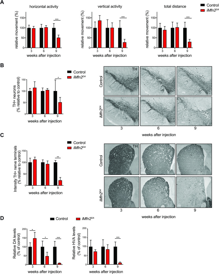
Fig 1. Tamoxifen-injected iMfn2DA mice show impaired locomotion and DA neurodegeneration.
Control and iMfn2DA mice were analyzed at 3, 6 and 9 weeks after tamoxifen or vehicle injection. (A) Spontaneous motor activity (horizontal activity, vertical activity and total distance) was measured in open field. ***p< 0.001, n>14. (B-C) Representative images of TH-like immunoreactivity in section from midbrain (Scale bars: 100 μm) and striatum (Scale bars: 200 μm), right panels. Quantification of TH-positive DA neurons in the midbrain and TH-immunoreactive nerve terminals in the striatum, left panels. ***p< 0.001 n = 3. (D) Analysis of DA and HVA levels in the striatum. Data are shown as mean ± SD. ***p<0.001 n≥5.

