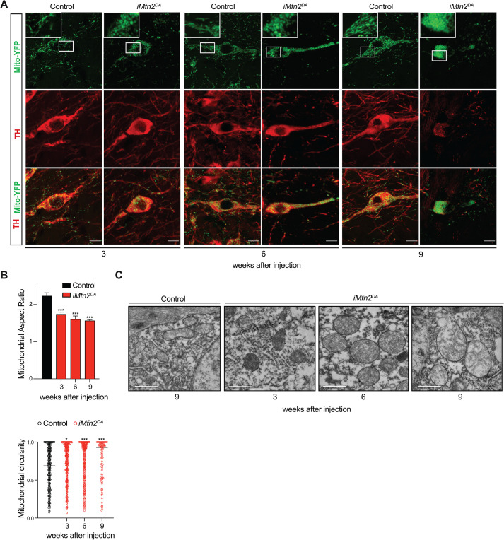
Fig 2. Loss of Mfn2 in adult DA neurons affects mitochondrial morphology and cristae structure.
Analyses were performed 3, 6, and 9 weeks after tamoxifen injection in control and iMfn2DA mice. (A) Representative confocal microscopy images of YFP-labelled mitochondria (green) in TH immunoreactive neurons (red) (Scale bars: 10 μm). (B) Quantification of aspect ratio and mitochondrial circularity from confocal images in TH+ DA neurons. AR data are shown as mean ± SD and circularity data as median of individual mitochondria. ***p< 0.00, n = 3 for each genotype. (C) Representative electron microscopy images of mitochondrial cristae structure (Scale bars: 1 μm).

