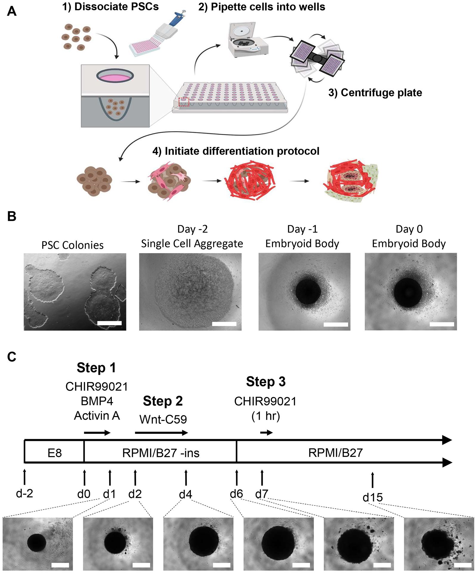
Figure 1: Embryoid body generation and heart organoid differentiation steps.

(A) (1–2) Dissociated cells are seeded into wells of a 96-well ultra-low attachment plate via a multichannel pipette. (3) The 96-well plate is then centrifuged, which allows the cells to aggregate in the center. (4) Over time, following the addition of growth factors and pathway modulators, the embryoid body begins to differentiate into several cardiac lineages and form spatially and physiologically relevant distinct cell populations surrounding internal microchambers. (B) Representative images of the progression of embryoid body generation, beginning with 2-dimensional iPSC culture (left) and ending with a Day 0 embryoid body (right); scale bar = 500 μm. (C) Summary of human heart organoid differentiation protocol, including chemical pathway modulators and inhibitors with respective timepoints, durations, and developing organoid images under light microscopy from day 1 to day 15; scale bar = 500 μm.
