
FIGURE 3.
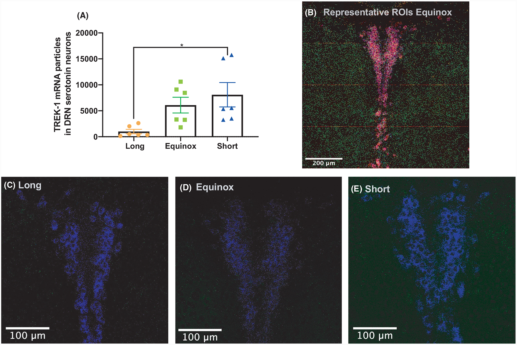
mRNA levels of Kcnk2 (TREK-1) in Tph2 and Pet-1-positive neurons are decreased in Long photoperiod mice. Quantification of colocalized Kcnk2, Tph2, and Pet-1 in the dorsal raphe. (A) Colocalized transcript detection of Kcnk2 in c3hf++ mice raised in long equinox or short photoperiods revealed a significant decrease of the transcript in serotonin neurons of long photoperiod mice (one-way ANOVA, F = 4.991, P = .0218*). Tukey’s post hoc multiple comparison revealed significant differences between Long and Short photoperiod cohorts. (B) Representative image of c3hf++ dorsal raphe from a mouse from the Equinox photoperiod, showing quantification of colocalized transcripts. The image was made from 16 captures at 40×. Kcnk2 (green), Tph2 (red), and Pet-1 (blue), colocalization is shown of Pet-1 and Tph2 is in purple, Kcnk2 colocalization is in either turquoise or yellow. Representative images of Kcnk2 (green) and Pet-1 (blue) signal from (C) Long, (D) Equinox, and (E) Short photoperiods. The brightness of Kcnk2 signaling for panels C, D, and E was increased by 20% in each image shown for visualization purposes.