Figure 5.

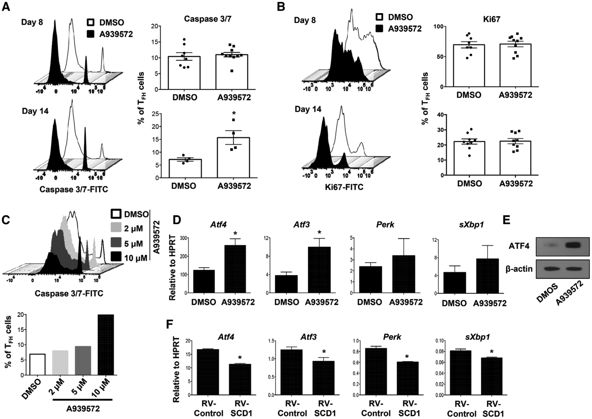
SCD inhibition promotes TFH apoptosis and ER stress gene expression. (A) Caspase 3/7 activity and (B) proliferation (indicated by Ki67 staining) of splenic TFH cells from the mice immunized with X31 virus and treated with SCD inhibitor at 8 and 14 d.p.i. (C) Naïve CD4+ T cells were stimulated under TFH polarization condition and treated with increased doses of SCD inhibitor, active caspase 3/7+ cells were measured at day 4 postactivation. (D) The expression of ER stress-related genes in CD4+ T cells under TFH polarization with or without SCD inhibitor (10 μM) was determined by qRT-PCR. (E) ATF4 protein level in CD4+ T cells under TFH polarization with or without SCD inhibitor. (F) The expression of ER stress-related genes in CD4+ T cells under TFH polarization with or without SCD1 transduction was determined by qRT-PCR. Combine data (A and B) are obtained from two independent experiments (two to five mice per group). Representative results (C and E) are shown from two independent experiments (two mice per group). Results are given as mean ± SEM with unpaired t-test: *p < 0.05.
